

Discover how photobiomodulation (PBM) may offer a novel approach to treating Myasthenia Gravis (MG). Explore its mechanisms in modulating immune responses, enhancing mitochondrial function, and potentially regenerating neuromuscular junctions


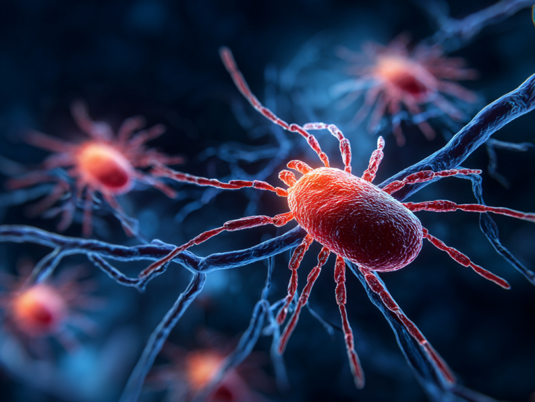
Myasthenia gravis (MG) is a chronic autoimmune disease characterized by skeletal muscle weakness and fatigability. The core pathology involves dysfunction of the neuromuscular junction (NMJ)—the critical communication hub between nerve and muscle. To explore the potential of emerging therapies like photobiomodulation (PBM), we must first understand the complex immunopathological mechanisms underlying MG.
The neuromuscular junction is a highly specialized synaptic structure formed between motor neuron terminals and muscle fiber membranes, serving as the key node for voluntary motor control. Its function depends on precisely coordinated physiological processes.
When a nerve impulse reaches the motor neuron axon terminal, it triggers the opening of voltage-gated calcium channels in the presynaptic membrane. Calcium influx promotes fusion of acetylcholine (ACh)-containing synaptic vesicles with the presynaptic membrane, releasing ACh into the synaptic cleft.
Released ACh molecules diffuse across the synaptic cleft and bind to nicotinic acetylcholine receptors (nAChRs) on the postsynaptic membrane. This binding causes AChR ion channels to open, allowing sodium influx and generating a depolarization potential known as the endplate potential (EPP). The small potential changes caused by individual synaptic vesicles are called miniature endplate potentials (mEPPs). When sufficient AChRs are activated and EPP amplitude reaches threshold, voltage-gated sodium channels in the muscle fiber membrane open, producing a propagating muscle action potential that ultimately triggers muscle contraction.
NMJ structural stability and functional integrity depend on the coordinated action of multiple key proteins. Among these, muscle-specific kinase (MuSK) and lipoprotein-related protein 4 (LRP4) are particularly important. LRP4 binds to neuron-released agrin, activating MuSK and initiating downstream signaling pathways that promote AChR clustering and stabilization at the postsynaptic membrane, forming high-density receptor clusters that ensure efficient neuromuscular transmission.
MG is a典型 B-cell-mediated autoimmune disease whose pathogenic core involves the body's immune system producing abnormal autoantibodies that attack key protein components of the NMJ. Based on different antibody targets, MG can be divided into several major subtypes, which show significant differences in immunopathology, clinical presentation, and treatment response.
AChR-MG: This is the most common subtype, accounting for approximately 85% of all MG patients. Patients develop autoantibodies against AChR, primarily IgG1 and IgG3 subclasses that effectively activate the complement system and serve as the main drivers of NMJ damage.
MuSK-MG: In some AChR antibody-negative patients, antibodies against MuSK can be detected. These antibodies are primarily IgG4 subclass. Unlike AChR antibodies, IgG4 antibodies typically do not activate the classical complement pathway. Their pathogenic mechanism mainly involves directly interfering with MuSK protein function, disrupting AChR cluster formation and maintenance, leading to NMJ structural and functional breakdown.
LRP4-MG: This is another less common subtype with LRP4 protein as the antibody target. LRP4 is a key component of the agrin-LRP4-MuSK signaling pathway, and anti-LRP4 antibodies interfere with normal pathway function, affecting NMJ formation and stability.
Different MG subtypes have fundamentally different pathogenic mechanisms that directly affect NMJ structure and function.
In AChR-MG, antibodies cause neuromuscular transmission failure through three main mechanisms:
In contrast, MuSK-MG has a completely different pathological mechanism. Its pathogenic IgG4 antibodies do not activate complement but functionally inhibit MuSK tyrosine kinase activity, leading to AChR cluster dissolution and interruption of NMJ maintenance signals. This not only affects receptor distribution but may also cause deeper muscle cell structural abnormalities, such as significant mitochondrial abnormalities observed in muscle biopsies from MuSK-MG patients.
The thymus plays a central role in AChR-MG pathogenesis. In approximately 70% of early-onset AChR-MG patients, the thymus shows follicular hyperplasia, forming structures similar to germinal centers in peripheral lymphoid organs. In another ~10% of patients, thymomas (usually benign) are present.
The scientific community widely believes that the abnormal thymus is the "source" of pathogenic anti-AChR antibody production. Within the thymus, helper T cells are abnormally activated, helping B cells differentiate into plasma cells and produce large amounts of anti-AChR autoantibodies. Myoid cells within the thymus express AChR on their surface and may serve as the initial source of self-antigen.
Thymectomy can significantly improve symptoms in many AChR-MG patients and may even reduce the need for immunosuppressants, providing strong clinical evidence for the thymus's central role in the disease. However, thymic abnormalities are uncommon in MuSK-MG patients, indicating weaker association between their pathogenesis and the thymus.
MG's hallmark symptom is fluctuating, easily fatigable skeletal muscle weakness that worsens with muscle use and improves with rest. Symptoms are typically most severe at the end of the day or after prolonged activity.
The clinical presentation spectrum is broad:
Ocular MG: Usually the initial symptom of MG, affecting approximately two-thirds of patients at onset. Main manifestations include ptosis and diplopia. Ocular MG may remain confined to the eyes, but in about half of patients, symptoms progress to generalized form within a few years.
Bulbar weakness: Affects facial, jaw, and throat muscles, causing dysarthria (slurred or nasal speech), dysphagia, and chewing weakness. Patients may easily choke while eating or experience liquid reflux from the nose.
Generalized MG: Weakness affects limb and trunk muscles, causing difficulty lifting arms, climbing stairs, or holding up the head.
The most serious complication of MG is myasthenic crisis, a life-threatening emergency. When respiratory muscle weakness (diaphragm and intercostal muscles) develops to the point where effective ventilation cannot be maintained, crisis occurs, requiring immediate intubation and mechanical ventilatory support. Common triggers include infection, surgery, psychological stress, or certain medications.
Additionally, various factors can worsen MG symptoms, including fatigue, illness or infection, surgery, psychological stress, pregnancy, menstrual cycles, and certain medications (such as β-blockers, certain antibiotics, and anesthetics). Notably, heat is also a recognized exacerbating factor—patients experience significantly worsened symptoms in hot environments or during fever.
Understanding MG's "fatigability" is crucial. This is not subjective tiredness but an objective electrophysiological phenomenon—progressive failure of neuromuscular transmission under repetitive nerve stimulation. The physiological basis is reduction of the NMJ "safety factor." In healthy individuals, the amount of ACh released from the presynaptic membrane far exceeds the minimum needed to trigger muscle action potentials—this redundancy is the safety factor. In MG patients, due to reduced AChR numbers or impaired function, this safety factor is severely eroded. When nerves fire repetitively at high frequency, ACh release naturally shows slight physiological decline. In healthy people, the large safety factor makes this decline inconsequential. But in MG patients, this small decline is sufficient to drop EPP below threshold, causing neuromuscular transmission failure and clinically manifesting as post-activity weakness.
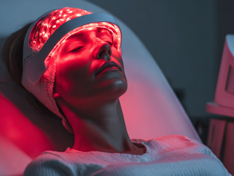
Photobiomodulation (PBM), formerly known as low-level laser therapy (LLLT), is a non-invasive treatment method that uses specific wavelengths of light to modulate cellular function, reduce inflammation, and promote tissue repair. To construct a scientific hypothesis for treating MG, we must first analyze how light energy is absorbed by the body and converted into beneficial biological effects.
PBM uses non-thermal doses of light, typically employing red light (~620-700 nm) and near-infrared light (NIR, ~700-1100 nm) wavelengths. These wavelengths are widely used because they fall within the "therapeutic optical window" of biological tissues. In this window, major light-absorbing substances in blood (hemoglobin) and skin (melanin) have relatively low absorption rates, allowing deeper light penetration to reach target cells and organelles.
PBM's biological effects begin when photons are absorbed by intracellular endogenous chromophores—natural light-absorbing molecules within cells. After photon energy absorption, these molecules undergo photochemical and photophysical reactions that initiate downstream cellular signaling cascades.
Mitochondria are the core organelles for PBM action, with a key enzyme serving as PBM's primary target.
Primary chromophore: The widely accepted primary chromophore for red and near-infrared light is cytochrome C oxidase (CCO), complex IV of the mitochondrial electron transport chain (ETC). CCO is the terminal enzyme of cellular respiration, responsible for transferring electrons from cytochrome c to oxygen, generating water.
Nitric oxide (NO) photodissociation: A widely accepted core hypothesis for how PBM activates CCO involves nitric oxide (NO) photodissociation. Under physiological or pathological conditions, NO can non-covalently bind to CCO's active center (heme and copper centers), competing with oxygen for binding sites and thereby inhibiting ETC activity and slowing cellular respiration. When specific wavelength photons are absorbed by CCO, their energy is sufficient to break NO-CCO binding, releasing NO. This clears the way for oxygen to rebind to CCO, rapidly restoring or even enhancing electron transport chain activity.
Increased ATP production: Enhanced CCO activity directly improves mitochondrial respiration and oxidative phosphorylation efficiency, leading to significantly increased adenosine triphosphate (ATP) synthesis. ATP is the cell's "energy currency," and increased production provides more abundant power for all energy-consuming cellular activities (ion pump operation, protein synthesis, cell repair, and muscle contraction).
Reactive oxygen species (ROS) regulation: PBM's regulation of reactive oxygen species (ROS) shows sophisticated, cell-state-dependent duality. In healthy, non-stressed cells, PBM can induce a brief, low-level ROS burst that serves as an important signaling event, activating cellular protection and repair-related signaling pathways. However, in cells already under oxidative stress (such as in inflamed or injured tissues), PBM's primary effect is to reduce pathological ROS overproduction and upregulate the cell's own antioxidant defense systems. Therefore, PBM doesn't simply "produce ROS" but acts as a regulator, pushing the cellular oxidative-reductive environment toward homeostasis.
One of PBM's most well-known and thoroughly researched effects is its powerful anti-inflammatory action, achieved through multiple pathways:
Cytokine profile transformation: PBM effectively modulates immune cell cytokine secretion profiles. Extensive research confirms that PBM can downregulate production of multiple pro-inflammatory cytokines such as tumor necrosis factor-α (TNF-α), interleukin-1β (IL-1β), and interleukin-6 (IL-6)—key molecules that drive and maintain chronic inflammation.
Simultaneously, PBM promotes secretion of anti-inflammatory cytokines such as interleukin-10 (IL-10) and transforming growth factor-β (TGF-β). This shift from pro-inflammatory to anti-inflammatory helps suppress excessive immune responses and restore tissue homeostasis.
NF-κB pathway regulation: Nuclear factor-κB (NF-κB) is a core transcription factor regulating inflammatory responses. PBM's regulation of NF-κB is also state-dependent. In resting cells, PBM-induced brief ROS bursts can activate NF-κB, initiating cellular protection programs. However, in already-activated inflammatory cells (such as LPS-stimulated macrophages), PBM shows inhibitory effects on NF-κB activity, blocking inflammatory cascade amplification at its source.
PBM's immunomodulatory effects extend beyond local inflammatory control to profoundly influence the adaptive immune system.
T-cell regulation: PBM can affect various immune cells including macrophages, dendritic cells, and T cells. Research shows PBM can modulate T-cell subset balance—for example, in allergic diseases, it can promote immune response shifts from Th2-dominated to more balanced Th1/Th2 states.
Promoting regulatory T cells (Tregs): For autoimmune diseases, one of PBM's most critical immunomodulatory mechanisms is its potential to promote regulatory T cell (Treg) differentiation and function. Tregs are the key "brake" system maintaining immune tolerance and suppressing autoreactive lymphocytes. By enhancing Treg function, PBM may help suppress the abnormal autoimmune responses causing MG.
Systemic effects: A striking phenomenon is PBM's systemic effects. Local body area illumination can produce beneficial biological effects in distant tissues and organs. The mechanism isn't fully understood but may involve circulating immune cells being modulated in illuminated areas then circulating systemically, or beneficial signaling molecules (cytokines, growth factors) induced by illumination being released into blood circulation, affecting the entire body. This concept is crucial for understanding PBM's potential in treating systemic diseases like MG.
A fundamental principle in PBM research is the biphasic dose-response, also known as the Arndt-Schulz law. This means:
This principle emphasizes that "more is not necessarily better." Therefore, determining optimal treatment parameters (wavelength, power density, illumination time, frequency) for any specific disease (including MG) is crucial for achieving clinical efficacy and ensuring safety—a core issue that must be addressed in clinical trial design.
This section is the core synthesis of our report, connecting MG pathophysiology (Section 1) with PBM mechanisms (Section 2) to construct a solid, evidence-based hypothesis explaining why PBM might become a viable MG therapy. We'll critically evaluate the currently very limited direct evidence and draw insights from related disease research.
Currently, published evidence directly applying PBM to MG treatment is extremely scarce, primarily concentrated in a 2022 case report describing a 59-year-old male AChR antibody-positive MG patient whose condition responded poorly to conventional treatments including acetylcholinesterase inhibitors and immunosuppressants—classified as refractory MG.
Intervention: The patient received intravascular laser irradiation of blood (ILIB) therapy—a systemic PBM treatment involving fiber optic insertion into veins for direct low-energy laser illumination of circulating blood.
Clinical outcomes: After three ILIB treatment courses, the patient's clinical symptoms improved significantly. The MG-ADL score (assessing daily activities) decreased from 17/24 pre-treatment to 3/24; the more objective quantitative MG (QMG) score also dropped from 32/39 to 9/39. The patient's diplopia, ptosis, and limb weakness all showed dramatic improvement.
Imaging evidence: Brain SPECT scans performed before and after ILIB treatment showed significantly increased cerebral blood flow perfusion in the frontal lobe and anterior cingulate cortex post-treatment, with this objective imaging improvement correlating with clinical symptom improvement.
Critical assessment:
Despite these limitations, this case report's value lies in first demonstrating PBM's feasibility for MG application and providing a strong signal worthy of further systematic study.
Based on PBM's known multiple biological effects, we can construct a comprehensive hypothesis explaining why it might have therapeutic effects on MG.
Hypothesis: Systemic PBM (such as ILIB or whole-body illumination) can modulate circulating immune cell function, rebuild immune tolerance, and thereby treat MG at its source.
Mechanism: MG's core is uncontrolled autoimmune response. PBM has been proven to suppress pro-inflammatory cytokine (TNF-α, IL-6) production while promoting anti-inflammatory cytokine (IL-10) secretion, effectively "cooling" the inflammatory environment driving autoimmune cycles. More specifically, PBM has potential to promote regulatory T cell (Treg) numbers and function. Tregs are key to suppressing autoreactive T and B cells. If PBM can enhance Tregs' suppressive function in patients, it could directly inhibit pathogenic antibody-producing B cell lineages, achieving comprehensive treatment effects.
Hypothesis: Local or systemic PBM can directly improve muscle cell energy metabolism, combating MG's characteristic muscle fatigability.
Mechanism: MG patients' muscle tissues, particularly in MuSK-MG patients, show mitochondrial abnormalities. PBM's core target is mitochondria, significantly increasing ATP production through CCO activation. This increased energy supply may provide multiple benefits: enhancing damaged NMJ compensatory capacity and improving remaining AChR work efficiency; providing more energy for ACh synthesis and release—high-energy processes; directly supporting muscle contraction processes, improving muscle endurance and reducing post-activity weakness symptoms. Additionally, PBM can reduce oxidative stress associated with muscle damage and inflammation, protecting muscle cells from further harm.
Hypothesis: PBM may possess capabilities to promote damaged NMJ structural repair and regeneration.
Mechanism (more speculative): While direct evidence is limited, the potential is enormous. In other neural or muscle injury models, PBM has shown tissue repair and regeneration promotion abilities. One study of local anesthetic-induced nerve damage found that 904 nm laser PBM treatment could significantly increase AChR ε-subunit expression. Another study noted that PBM could temporarily maintain AChR levels in denervated muscles. These findings, while from non-MG models, are highly inspirational. They first suggest PBM may possess potential to directly influence AChR expression. If PBM could stimulate new AChR synthesis in MG patients or protect existing AChRs from immune attack destruction, this would directly counter the disease's core pathological elements, fundamentally improving neuromuscular transmission.
Comprehensively viewed, PBM's potential benefits for MG are multifaceted. It may address the "cause" (autoimmunity) through immune modulation while also alleviating "symptoms" (muscle fatigue) through mitochondrial function enhancement. This unique "dual approach" potential distinguishes it from most existing single-target therapies and constitutes a strong theoretical foundation for researching its MG applications.
Although PBM research in MG is in its infancy, its applications in other diseases with similar pathological features provide valuable indirect evidence.
Fatigue-related diseases: In fibromyalgia and chronic fatigue syndrome—diseases characterized primarily by severe fatigue—PBM has shown efficacy in reducing fatigue and improving physical capacity. These diseases share debilitating fatigue symptoms with MG, making their treatment experiences relevant.
Other autoimmune diseases: PBM is being actively researched for treating rheumatoid arthritis (RA), multiple sclerosis (MS), and Hashimoto's thyroiditis. Studies show PBM can reduce autoantibody levels, modulate inflammatory responses, and improve clinical symptoms. These studies confirm PBM's potential as an immunomodulatory tool, providing collateral evidence for MG applications.
Neuroinflammation and neurodegenerative diseases: In traumatic brain injury (TBI) and stroke models, PBM has been proven to reduce neuroinflammation and improve cerebral blood flow. This aligns with the SPECT cerebral blood flow improvement observed in the Lan et al. case report, suggesting PBM's MG benefits may extend beyond peripheral muscles to central nervous system modulation.
This series of indirect evidence collectively points to one conclusion: PBM's biological effects are highly compatible with the pathological mechanisms of multiple chronic, inflammatory, and autoimmune diseases. Particularly, its demonstrated potential in MG case reports is not an isolated phenomenon but a manifestation of its broad therapeutic effects in this specific disease.
Translating a theoretically promising therapy into safe, effective clinical practice requires overcoming numerous challenges. This section explores practical issues in MG clinical applications of PBM, including application methods and safety considerations, while proposing research pathways for the future.
Based on PBM's mechanisms of action and MG's disease characteristics, future potential clinical application modalities include:
Systemic applications:
Local applications: Direct PBM device application to symptomatic muscle groups such as neck, shoulders, and limbs. This approach aims to rapidly relieve symptoms by enhancing local muscle ATP generation and reducing local inflammation. Advantages include strong targeting and simple operation. Disadvantages may include inability to address underlying systemic autoimmune issues.
Hybrid modalities: A highly promising strategy combining systemic PBM (such as full-body light therapy beds) to modulate overall immune status while providing additional local PBM treatment to particularly symptomatic muscle groups, seeking synergistic effects of systemic modulation and local enhancement.
While PBM is generally considered safe, specific risks must be considered when applying it to this special MG patient population.
General contraindications: PBM should not be directly used on known malignant tumor sites (unless permitted by physicians for palliative treatment), eyes (specifically laser sources), or pregnant women's uterine areas.
Heat sensitivity paradox: This is a crucial consideration when applying PBM to MG patients.
Thyroid considerations: MG patients often have other autoimmune diseases, including thyroid diseases (such as Hashimoto's thyroiditis). PBM has been proven to affect thyroid function. Therefore, when treating neck areas, caution should be exercised to avoid direct thyroid illumination, or thyroid function should be closely monitored before and after treatment.
Moving from hopeful hypothesis to widely accepted therapy requires rigorous scientific validation.
Preclinical research: The most urgent next step is validating PBM efficacy in established experimental autoimmune myasthenia gravis (EAMG) animal models. EAMG models can well simulate key pathological features of human MG, including autoantibody production and NMJ damage. In these models, researchers can thoroughly explore PBM mechanisms under controlled conditions: Can it reduce anti-AChR antibody titers? Can it increase Treg numbers in spleen and lymph nodes? Can it protect NMJ ultrastructure? Can it increase muscle tissue ATP levels? Only with positive answers in animal models can we provide solid scientific basis and ethical support for human trials.
Phase I/II human clinical trials:
Although PBM's application prospects in MG are broad, many key questions urgently need answers:
The SPECT imaging improvement revealing increased cerebral blood flow in the Lan et al. case report is a very important clue. It suggests PBM's MG benefits may extend beyond peripheral muscles and immune systems. MG fatigue has central components, and PBM has shown cerebral blood flow improvement and neuroprotective effects in other neurological diseases. This raises a more comprehensive "neuro-immune-muscle" three-dimensional action hypothesis: PBM may reduce the "central effort sensation" required to generate movement by improving central motor pathway efficiency and reducing neuroinflammation, thereby alleviating fatigue from another dimension. This "neuro-centric" hypothesis opens new directions for future research.
Research synthesis: Myasthenia gravis is a complex autoimmune disease whose core pathology involves immune system attacks on neuromuscular junctions, resulting in clinical syndromes characterized by fluctuating, easily fatigable muscle weakness. Photobiomodulation, as an emerging therapeutic approach, has extensive research confirming its mechanisms of enhancing energy metabolism at the cellular level, suppressing inflammation at the tissue level, and modulating immune responses at the systemic level.
Core argument: This report systematically demonstrates that using PBM as a novel MG therapy has strong, multi-level theoretical foundations. PBM's mechanisms of action are highly compatible with MG's core pathological elements, potentially achieving unique dual benefits: suppressing underlying autoimmune causes through immune modulation while directly alleviating muscle weakness—the primary symptom—through enhanced bioenergetic metabolism.
Evidence status: However, this strong theoretical foundation currently has support only from one highly inspirational but isolated human case report, plus substantial indirect evidence from related disease fields. Between PBM's enormous theoretical potential for treating MG and definitive clinical validation exists a significant gap that urgently needs filling.
Final statement: Photobiomodulation represents a non-pharmacological, potentially transformative frontier direction in myasthenia gravis treatment. It is not currently a mature therapeutic method but rather an urgently needed, compelling scientific hypothesis awaiting validation. Future research must systematically test whether its theoretical promises can translate into clinically meaningful benefits for patients through rigorous preclinical animal models and carefully designed randomized controlled human trials. This exploratory path may bring


